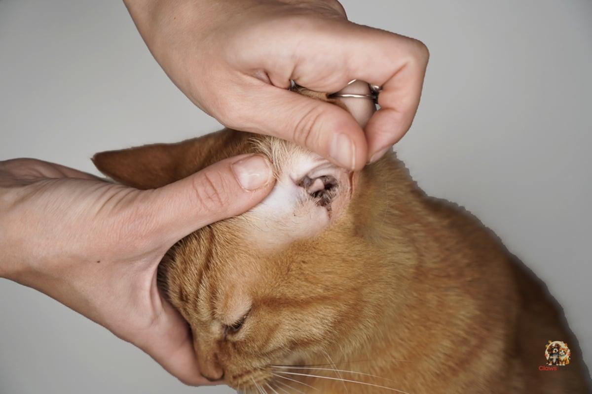
Today, in the company of Dr. Cattaneo, a veterinarian at the Pineta Veterinary Clinic, we will talk about dermatophytosis, superficial and contagious infections caused by microscopic fungi, visible only under a microscope. The main component of fungi are hyphae—branched tubular formations divided by transverse septa, which define them as filamentous or mycelial microorganisms. This video will focus on the microscopic fungi of medical interest, known as dermatophytes.
Yeasts are non-filamentous microscopic fungi composed of unicellular elements that reproduce by budding. Examples of yeasts include Malassezia and Candida. But today, we are talking about dermatophytes.
How do dermatophytosis manifest, and what are the most common ones?
Dermatophytosis is contagious, superficial infections caused by pathogenic fungi. These fungi are pathogenic because dermatophytes cannot multiply in the external environment, but only on the animal's skin. They can spread from a sick animal to a healthy one or through a contaminated environment, making them contagious. They mostly cause superficial infections because they affect the keratinized cells of the epidermis, nails, and hair. They cannot multiply in the environment, but their spores can last a long time.
These animal dermatophytoses are transmissible to humans, posing a significant public health concern. Microsporum canis is the most common dermatophyte found in dogs and cats, with cats being the most common source of infection. Transmission can occur through direct contact or via occasional spore carriers like shoe soles, clothes, or air conditioning, especially in high-contamination areas like kennels, catteries, and farms. Veterinary facilities, like any place frequented by many animals, can also potentially be a source of infection.
Other dermatophyte species that can cause disease in our pets include Trichophyton mentagrophytes, whose reservoir includes rodents and lagomorphs such as hares and wild rabbits, and Microsporum persicolor, which affects not only dogs but also small rodents.
How can dermatophytosis be clinically recognized?
Depending on each organism's response to the infection, the clinical manifestations of dermatophytosis can vary greatly and overlap with the clinical manifestations of numerous dermatological diseases, making differential diagnosis necessary.
Once contracted, the infection can progress differently depending on whether it affects a dog or a cat. The breed, coat characteristics, skin, immune status, age, overall health conditions, environmental and hygienic conditions, and the infectious load all play a role. The time before clinical lesions appear is typically 1-2 weeks.
In dogs, the most commonly observed lesions are hairless, round-shaped areas, single or multiple (multifocal), with a diameter of 1-2 centimeters. If the animal's immune response causes inflammation, redness (erythema) and scales of varying sizes, from white to grayish, may develop.
The muzzle, back of the nose, ears, and paws—areas more exposed to other animals or rubbing—have the most lesions. Depending on the subject’s receptivity, a centrifugal expansion may occur, and the lesions tend to lose their initial defined margins, expand, and merge. Paradoxically, the suspicion of diagnosis can become more difficult when the infection is older due to a loss of the initial identifiable appearance.
The individual response is extremely variable, so the appearance of papules and pustules can be complicated by the presence of neutrophil granulocytes or exaggerated immune responses that alter the clinical picture, making diagnosis more difficult.
Are there any breeds more susceptible to the infection?
Certain dog breeds, like terriers and poodles, seem to be more sensitive to the infection and predisposed to developing generalized forms. For cats, Persian cats and generally long-haired breeds appear to be predisposed to developing the disease.
As mentioned, the immune system plays an important role: puppies, kittens, elderly, or weakened animals are at higher risk. As a result, any debilitating illness that reduces immune defenses makes dogs and cats more susceptible to dermatophytosis. Therefore, underlying pathologies must be investigated and treated before starting specific therapies for fungal infections.
Excessive washing with aggressive shampoos also reduces the skin's natural defenses. Parasitic infections, pruritic dermatitis caused by other factors, or other types of dermatitis can create entry points for dermatophytes.
Let's not forget climatic conditions: humidity or excessive heat are predisposing factors. Stress, overcrowding in kennels, boarding houses, or training camps in relation to the presence and quantity of spores also play a role in the development of dermatophytosis.
How is the diagnosis made?
Diagnosis is made through various methods. First of all, in case of clinical suspicion, a Wood’s lamp is used because hairs invaded by M. canis (the most common dermatophyte) may show a yellow-green fluorescence under ultraviolet light. But fluorescence alone isn't enough to make a diagnosis. The diagnosis must also be confirmed by looking at the fluorescent hairs under a microscope that were collected during the lamp examination and by growing the fluorescent hairs in a culture medium that is specifically made for dermatophytes.
Even in the absence of fluorescence under the Wood’s lamp, it is still necessary to collect and microscopically examine the hairs from the center of the alopecic lesion, as most pathogenic dermatophytes do not fluoresce under the Wood’s lamp. Hairs should be collected gently, respecting the direction of growth to avoid breaking the hair shaft and losing the infected portion.
Fungal culture on specific media for dermatophytes (DTM) is still a reliable technique for confirming dermatophytosis. Samples are obtained by plucking hairs under a Wood's lamp at suspicious lesions, or by brushing the coat with a sterile toothbrush. Scales should also be collected and sown. The plates with the collected material are incubated at a controlled temperature (25 °C) for about two to four weeks.
The plates are observed daily to detect the color change characteristic of dermatophyte colony growth. If this occurs, the dermatophyte is then identified and typed.
How is dermatophytosis treated?
We use complementary topical and systemic antifungal treatments once we confirm the infection. Systemic therapy treats the hair follicle, while topical products eliminate the spores on the hair surface.
Conventional systemic treatment is based on oral antifungals administered for the necessary duration. The owner must discuss the decision to use topical therapy, which also aims to reduce the risk of environmental contamination. Bathing or spongeing the entire coat of the infected animal requires skill and motivation; local treatments should be performed at least twice a week.
It is important to remember that infected and uninfected animals must be completely separated, as this is a zoonosis—a disease that can be transmitted from animals to humans!
Dermatophytosis: how can it be prevented?
The best method is to avoid contact with infected animals or environments, as dogs and cats can contract the infection at any time in their lives.
Since there are infected individuals who do not yet show clinical signs or who do not necessarily show evident clinical signs, it is not always possible to avoid the risk of infection. In dog and cat breeding facilities and animal shelters, the introduction of an infected animal is the biggest risk, so careful clinical examination of each new individual is essential.
It is also important to implement procedures to reduce the risks of other diseases: before introducing new animals, they should be vaccinated, dewormed, treated for external parasites, examined with a Wood’s lamp, and quarantined until the culture result is available. These procedures are not always feasible due to space, cost, and time constraints, which increase the risk of infection.
What should owners of infected animals pay particular attention to?
The most important preventive measures for owners include paying special attention to personal hygiene due to the risk of being infected, having regular diagnostic tests performed, and following the prescribed treatments for their pet. It is also advisable to avoid direct contact between infected animals, contaminated environments, and children or individuals with immune problems. Veterinarians must inform those in contact with infected animals about the risks and provide behavioral guidelines to reduce the possibility of infection.
More: Dog Health | Cat Health


